Expert Urological Care & Minimally Invasive MIST Center in Seoul

Medical Guide
Life After Shock Wave Lithotripsy (ESWL): A Recovery Timeline
Learn what life after Shock Wave Lithotripsy (ESWL) looks like, including recovery milestones, discomfort, activity, and aftercare. Explore the timeline today.
Schedule a ConsultationWhat Is ESWL and Why Is It Used?
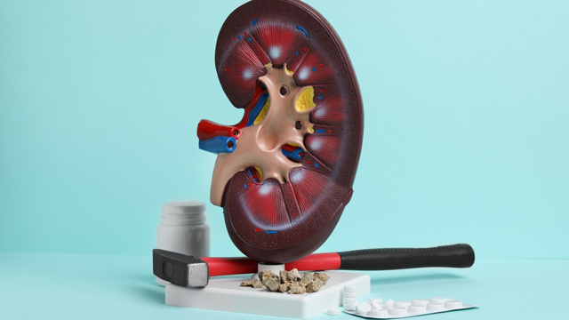
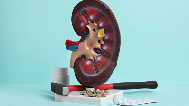
ESWL is a non-surgical treatment that uses focused shock waves, generated outside the body, to break kidney stones into smaller fragments. Once fragmented, these smaller pieces can pass through the urinary tract more easily, typically in urine, over the days and weeks following treatment.
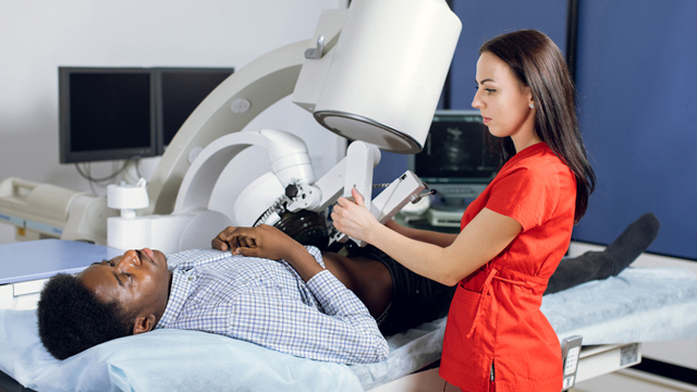
Unlike surgical stone removal, ESWL requires no incisions. The shock waves are aimed precisely at the stone using real-time imaging guidance — usually ultrasound or fluoroscopy (a type of moving X-ray) — allowing the surrounding kidney tissue to remain largely unaffected while the targeted stone absorbs the mechanical energy. Over repeated shock wave pulses, the stone's internal structure weakens and fractures along natural planes, eventually breaking into pieces small enough to be passed naturally.
This technology has been used clinically for several decades and remains one of the most widely performed treatments for kidney stones globally, precisely because it combines a reasonably high success rate with a low complication profile for appropriately selected patients.
How ESWL Compares to Other Stone Treatments
Kidney stones can be managed in several ways depending on size, location, and composition:
Observation and hydration — appropriate for very small stones (often under 4–5 mm) that are likely to pass on their own.
ESWL — a non-invasive option well suited to moderate-sized stones in favorable locations.
Ureteroscopy — a minimally invasive procedure using a thin scope passed through the urinary tract to directly visualize and remove or fragment the stone, often used for stones in the ureter or when ESWL is not suitable.
Percutaneous nephrolithotomy (PCNL) — a more invasive procedure reserved for larger or more complex kidney stones, involving a small incision in the back to access the kidney directly.
ESWL sits in a middle ground: more active than watchful waiting, but far less invasive than surgery. Its main appeal lies in the fact that it can often be performed without general anesthesia, without incisions, and typically on an outpatient basis — allowing patients to resume normal activities more quickly than after surgical alternatives.
Who Is a Good Candidate for ESWL?
Not every stone or every patient is suited to shock wave treatment. Stone-specific and patient-specific factors both play a role in determining candidacy. Generally, ESWL tends to work best for:
Stones roughly 4–20 mm in size
Stones located in the kidney or upper ureter
Stones with a composition that responds well to shock wave energy, such as calcium oxalate dihydrate or uric acid stones
Patients without significant bleeding disorders or uncontrolled infection
Patients who are not pregnant, since shock waves are not used during pregnancy
Patients with a body habitus that allows the stone to be clearly visualized and targeted with imaging
Larger stones, very hard stone types (such as certain cystine or calcium oxalate monohydrate stones), or stones lodged in difficult anatomical positions — for example, deep within the lower pole of the kidney, where gravity works against fragment clearance — may respond better to alternative treatments. Body weight and the distance between the skin surface and the stone can also affect how efficiently shock waves reach their target, which is why imaging evaluation beforehand is essential.
A thorough evaluation — including CT or ultrasound imaging, urinalysis, and in some cases prior stone analysis — helps determine whether ESWL is the right fit before treatment begins. This individualized assessment is a key part of avoiding unnecessary procedures and setting realistic expectations from the start.
What Happens During the Procedure?
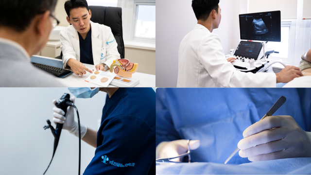
ESWL is typically performed as an outpatient procedure, meaning most patients go home the same day. While specifics can vary slightly by clinic and equipment, the general process includes:
Pre-procedure evaluation — Imaging confirms the stone's size, location, and density, and baseline labs may be reviewed to rule out active infection.
Positioning — The patient lies on a treatment table, and the shock wave device is aligned with the stone using real-time imaging guidance.
Pain management — Depending on the clinic's protocol and the patient's tolerance, mild sedation, local anesthesia, or oral analgesia may be used, since some patients feel a tapping or thumping sensation during treatment.
Shock wave delivery — A series of focused shock wave pulses is directed at the stone over the course of the session, gradually fracturing it. The number of pulses and treatment duration are adjusted based on stone response.
Monitoring — The physician monitors the treatment in real time, adjusting shock wave intensity and targeting as needed based on how the stone responds and how the patient tolerates the session.
Post-procedure observation — A short observation period follows to check for immediate complications before the patient is discharged.
Most sessions are completed within about 30 to 60 minutes, and many patients are able to return home the same day, often resuming light activities within 24 hours.
Life After ESWL: A Recovery Timeline
Recovery from ESWL is generally faster than recovery from surgical stone treatment, but it still follows a predictable pattern that patients should be aware of. Below is a general timeline; individual experiences can vary based on stone size, number of fragments, and overall health.
Day 1–2: Immediate Post-Procedure Period
It's common to experience mild discomfort, bruising over the treated skin area, or blood-tinged urine (hematuria) in the first day or two. This occurs because the shock waves pass through skin and soft tissue on their way to the stone. Drinking plenty of water — often at least 2 to 3 liters daily unless otherwise restricted — is usually recommended to help flush out stone fragments and reduce the risk of blockage.
Over-the-counter or prescribed pain relief may be used as directed by the treating physician; anti-inflammatory medications are sometimes preferred, though the physician will tailor the choice based on kidney function and any other health conditions. Light walking is often encouraged, as gentle movement can help facilitate fragment passage, while strenuous exercise is typically discouraged for the first few days.
Day 3–7: Stone Fragment Passage
This is typically when most stone fragments begin to pass. Some patients notice gritty material or small fragments in their urine — this is expected and is often a sign the treatment is working. Straining urine through a fine mesh or cloth, if instructed, can help capture fragments for stone composition analysis, which may guide future prevention strategies, including dietary changes or targeted medication.
Mild, intermittent cramping or flank discomfort can occur as fragments move through the ureter, sometimes described as similar to mild renal colic. Staying well hydrated and maintaining light activity generally supports this process. However, certain symptoms are not considered part of a normal recovery and should prompt immediate medical attention:
Sudden, severe pain that doesn't respond to prescribed medication
Fever or chills, which may indicate infection
Persistent nausea or vomiting
Inability to urinate, which may indicate an obstructing fragment
These warning signs can indicate complications such as a urinary tract infection or a fragment causing a blockage, both of which require prompt evaluation.
Week 2–4: Continued Clearance and Follow-Up Imaging
By this stage, most smaller fragments have typically passed. A follow-up visit with imaging — often ultrasound or a low-dose X-ray (KUB) — is usually scheduled to confirm that fragments have cleared and no significant residual stone remains. This follow-up is an important step, since some fragments may be too large to pass spontaneously even after successful fragmentation.
If larger fragments persist beyond this window, additional treatment — whether a repeat ESWL session, ureteroscopy, or another approach — may be discussed based on fragment size and the patient's symptoms. Steinstrasse, a rare complication in which a column of fragments becomes lodged in the ureter, is one reason follow-up imaging matters even in patients who feel well.
Beyond One Month: Long-Term Monitoring and Prevention
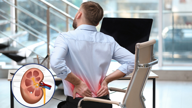
Once initial recovery is complete, attention shifts to prevention, since kidney stones have a meaningful recurrence rate — studies generally suggest a notable proportion of patients form another stone within five to ten years without preventive measures. Physicians often recommend:
Increased daily fluid intake, generally enough to produce pale-colored urine throughout the day
Dietary adjustments based on stone composition — for example, moderating oxalate-rich foods for calcium oxalate stones, or reducing purine-rich foods for uric acid stones
Attention to sodium intake, since excess salt can increase urinary calcium excretion
Periodic imaging follow-up, particularly for patients with a history of recurrent stones
Metabolic evaluation in select cases — including 24-hour urine testing — to identify underlying risk factors such as low urine volume, high calcium excretion, or low citrate levels
Safety Considerations and Realistic Expectations
ESWL is considered a safe procedure for appropriately selected patients, but like any medical treatment, it is not without limitations. Success rates vary depending on stone size, location, density, and composition, and some patients may require a second session or an alternative procedure if fragments do not clear as expected on the first attempt.
Less common risks can include significant bleeding around the kidney, persistent obstruction from larger fragments, or infection, though these occur in a minority of cases when patients are properly screened beforehand. Setting realistic expectations from the outset — rather than assuming a single treatment guarantees complete stone clearance — leads to better-informed decisions and less anxiety during recovery. Patients who understand that fragment passage can take days to weeks, and that follow-up imaging is a normal part of the process rather than a sign that something went wrong, tend to navigate recovery with more confidence.
A Patient-Centered Approach to Urological Care
At Straight Urology Clinic, located in Bukahyeon-dong, Seodaemun-gu, Seoul — just steps from Ahyeon Station on Line 2, with convenient access to Seoul Station, the Incheon Airport Railroad, and KTX — the treatment philosophy centers on accurate diagnosis and appropriately matched care, rather than one-size-fits-all treatment. Under the direction of Dr. Dong-hyun Lee, who was educated abroad and provides full consultations directly in English, international patients can discuss their diagnosis, treatment options, and recovery expectations without the need for interpretation.
The clinic's diagnostic approach — including ultrasound imaging, uroflowmetry, and laboratory testing — reflects the same evidence-based philosophy applied across its urological services: identifying the underlying cause of a patient's symptoms, explaining findings in clear, plain language, and recommending only the level of treatment that is clinically necessary. This philosophy of avoiding over-treatment while ensuring thorough diagnostic evaluation is central to how care decisions are made, whether for prostate conditions, female urological concerns, or general urinary symptoms.
Final Thoughts
Recovery after ESWL generally follows a predictable arc: mild discomfort and possible bruising in the first days, fragment passage over the following one to two weeks, and follow-up imaging within a month to confirm clearance. While most patients recover quickly and resume normal activities within a few days, staying alert to warning signs — persistent fever, severe pain, or inability to urinate — remains important throughout the process. Combined with attentive follow-up care and a personalized prevention plan tailored to stone composition and individual risk factors, ESWL can be an effective, minimally invasive option for many patients dealing with kidney stones.
This article is for general informational purposes and does not replace personalized medical advice. Patients should consult a qualified urologist to determine the most appropriate diagnostic and treatment approach for their specific condition.
More Articles
Continue reading clinical insights and recovery guides from Straight Urology Clinic.
Who Is a Candidate for REZUM Water Vapor Therapy
Learn who may be a candidate for REZUM Water Vapor Therapy, key eligibility factors, benefits, and what to expect. Explore your treatment options today.
Learn More
Why a Urologist's Case Volume Should Matter to You
Surgical case volume is linked to a urologist's experience but never guarantees your outcome. Learn to read reported urology procedure counts critically.
Learn More
What AUA Membership Means for Your Urologist Choice
Learn what American Urological Association Full Membership actually means, why it is not a board certification, and how to verify an overseas urologist.
Learn More